Are Dental X-Rays Safe at My Rockford Dentist?
Trust and transparency are a top priority for your oral health. Hence, if you would like to confirm whether your Rockford dentist’s dental X-rays are safe, it is not rubbish to be worried. Even though dental X-rays are an indispensable diagnostics tool, the fact that they expose patients to radiation is a legitimate issue. Now, let us explore facts, find out what is in reality and myth, and give you the power to choose whether your dental care is right based on facts.
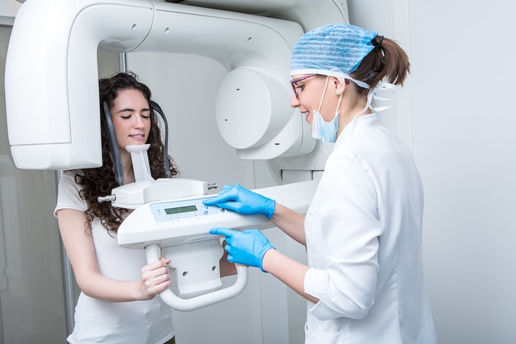
Understanding Dental X-Rays
Dental X-rays are not just simple pictures but tools that give us an exceptional view into the hidden part of the mouth where your teeth and jaws are. These crucial diagnostic tools unveil a wealth of information that might remain invisible to the naked eye during a routine exam, empowering dentists to: These critical diagnostic tools unveil a wealth of information that might remain invisible to the naked eye during a routine exam, empowering dentists to:
- Identify hidden threats: Cavity in the middle of the teeth, bone loss jeopardizing the stability of the teeth, and even early signs of infections can be detected by an X-ray so that intervention can be done immediately, preventing complications that may develop.
- Guide treatment decisions: Whether you need a simple filling or a more complex treatment, including installing a root canal or implant, detailed X-ray Images give dentists a plan and how to perform surgery.
- Monitor your oral health journey: Tracking tiny shifts in time through routine X-rays enables a dentist to see if the treatments are working correctly, spot any developing issues, and keep a watchful eye on your oral health as you grow up.
Exploring the Specialized Arsenal of X-ray Types:
Just like soldiers carry different tools for specific missions, dentists utilize various X-ray types tailored to uncover unique information:
- Bitewing X-rays: Have you ever imagined two opposing armies in the battle line? Bitewings grab not only the lower and the upper teeth in one area but also get deep through cavities hidden between them, especially among children susceptible to interdental decay patients.
- Periapical X-rays: Think of the detailed examination of a soldier by going through them with a magnifying glass. Periapical X-rays deliver comprehensive pictures of individual teeth, enabling dentists to make decisions regarding the health of your roots, detect any infection, and evaluate the bone density around each tooth.
- Panoramic X-rays: Consider a shot that includes everything from the lines of soldiers on different sides. Panoramic X-rays provide a comprehensive overall picture of your entire mouth; they show how the jaw develops, whether there are wisdom teeth and the actual layout.
Remember, you will take the type of dental x-ray according to your needs and what the dentist wants to know. Armed with the information in this article, you can go for your next visit to the dentist feeling empowered and self-aware, ready to work together with your dentist toward a healthy and radiant smile.
Safety of Dental X-Rays:
Modern dental care strives to create a safe environment for patients, and reducing the amount of radiation during X-rays is a significant aim. So, take a deep breath – here’s why you can face your subsequent X-ray with confidence: So, take a deep breath – here’s why you can meet your subsequent X-ray with confidence:
Shrinking Radiation Giants: Decrease Doses with Cutting-Edge Technology.
I bet you have seen those thick X-ray machines in movies or remembered the imposing X-ray machines of the senior dental offices! Just contemplate the panic they would have spread! Fret not; all those bellowing radiation-emitting colossi are replaced with new-age precision machines, thus decreasing exposure levels. Here’s how:
From Titanic to Speedboat: Drastically Reduced Radiation Doses:
The principal discrepancy depends on the radiation sources themselves. The predecessors to today’s X-ray machines included heavy and oversized filament bulbs that radiated quite strong and broad radiation beams. Picture this as if you are trying to focus a light similar in size to the car on a specific object by using the spotlight. Now, picture the arrow. The X-rays in modern dentistry are generated using solid-state technology, which produces the focused and collimated beam. Such precision-guided targeting gives rise to an almost 80-90% reduction in radiation emission levels compared to those experienced in earlier times. Thus, whereas the old machines made it like shining a flashlight all over your entire body, the current technology instead directs light only to the needed area, minimizing the exposure of the other regions.
Digital Revolution: Smaller Beams, Sharper Images:
Think of those old-fashioned grainy photos captured on film. They are the high-definition improvement in the field of dentistry. Including these systems allows for the images to be taken electronically using even smaller and focused beams. It could be likened to the difference between a standard-definition camera and a high-resolution one. On top of the higher quality and more detailed images, the smaller beam also helps to reduce radiation dose. Just imagine what it would be like if you reversed the work of a searchlight with a laser beam; that would be the power of digital technology!
Beyond Numbers: Real-World Examples for Clarity:
And let’s see from this what such a reduction looks like. The m Sv of radiation that results from each traditional bitewing X-ray of the past is 0.1. With modern technology, one bitewing x-ray now emits 0.01-0.04 mSv of radiation(a 90% reduction). Indeed, that is equivalent to the 2018 average background radiation exposure of 3.1 mSv in the U.S., which is only about 1/3200th of one average X-ray they receive in a year.
Thus, when you go for the dental x-ray next time, you will be pleased to know that the modern equipment is not the fish in the radiation– pond anymore. An analogy to their agile jets is a functional description. They are ridges of a set, delivering necessary information with minimum exposure, so your oral health journey makes safety and diagnostic accuracy the main priorities.
Precision Beats Scatter: Targeting Radiation for Minimal Exposure
Think of a shotgun with its large spread of bullets compared to a precision laser, which can get to a point only as wide as a pinhead. The benefit of digital X-rays over traditional film X-rays is the radiation exposure. Let’s delve deeper into this crucial aspect of safety: Let’s delve deeper into this vital aspect of safety:
Scattering: The Unwanted Guest at the Radiation Party:
X-rays worked like shotguns in the past, blasting their beams at wide angles to see the desirable image and affect the surrounding tissues. The “Scatter” effect brings under-exposed and over-exposed needlebeams to the overall dose. What’s the point in throwing confetti if it just falls off everywhere but mostly lands where you wanted it?
Digital Revolution: Pinpoint Accuracy Minimizes Unwanted Exposure:
And it went digital X-rays, which led to nothing but game changers in targeted imaging. These systems of the highest level employ the non-spread light, like lasers, that were applied to the desired field of view. This high precision dramatically minimizes “scatter” and, as a result, reduces the dose radiation that reaches un-intended tissues or organs. Try using a laser pointer instead of the confetti – perfect targeting and fewer hits on unintended targets.
Benefits Beyond Safety: Sharper Images for Better Diagnosis:
The benefit of digital X-rays lies in their specialized nature, which promotes safety and makes diagnostics much more accessible. Details since “scatter” inhibition become sharper and more precise. This enables dentists to find out the trivial and the slightest problems, such as early cavities or bone loss, with much more accuracy, which helps them make more accurate treatment plans. It is like comparing a hazy, defocused photo to a high-resolution one. The more clear details enable a better understanding, and, therefore, the more informed decisions can be made.
Real-World Example: Putting Precision into Perspective:
Take into account the typical panoramic X-ray radioscopy. The prior pattern would have left an extensive part of your head and neck at risk of irregular rays. On the other hand, the well-directed beam uses digital technologies that aim at the jaw and teeth, thus decreasing exposure to the fragile cells in the surrounding areas, such as the thyroid. This reduction of the scatter minimizes safety and is more tolerant to more frequent X-rays; hence, they can be done without much harm to monitor a particular case.
Remember, precision is key! Through this type of beam and fewer scattered ones, dental digital radiographs provide safety and allow you to receive more exact diagnoses and appropriate care for oral health. Hence, the next time you go to the clinic for an X-ray, you should feel relaxed and confident knowing that technology has your back, delivering safety and accuracy in a single focused beam.
Shielding Your Body’s Fortress: Protective Gear Minimizes Risk:
Imagine yourself immersed in the medieval world of a battlefield set in the distant past, but instead of knight’s armor, you have a high-tech shield to defend yourself. That’s your thyroid collars and lead aprons that your dentist wears while conducting an X-ray, turning them into your private bodyguards from the stray radiation field. Here’s how these shields minimize risk: Here’s how these shields reduce risk:
Lead Aprons: Your Vital Link of Protection to Shield from Sideshots Radiation.
Imagine this as an apron that is wide, powerful, and draped on your torso and lap. These aprons were made from a very high ray diffusion material, thus better protecting the organs by diverting the rays like the lungs, heart, and abdomen. Think about it as the barrier that suppresses the radiation, shelling it out into isolated places and reducing its ability to be dangerous.
Beyond the Obvious: Shielding Vulnerable Neighbors:
Even though the lead apron shields your central body, the other critical zone is also protected, the thyroid gland. The thyroid in your neck is a target organ due to its high affinity for iodine. This is when the thyroid collar comes in.t Lead or lead-equivalent material being its composition, it is a little shield when it gets small and targeted. Thus, it can prevent your thyroid from wandering radiation.
Beyond Armor: Minimizing Exposure Through Positioning:
Countershields are critical, but positioning can be equally important. Your dentist will properly set you while an X-ray is done and ensure that the beam is projected away from sensitive parts of the patient throughout. Imagine it as a move of strategic positioning towards the wall of your ‘fortress’ to keep you away from unnecessary contact.
Remember, It’s All About Minimizing Risk:
Although X-ray movies expose minimal radiation doses, each measure is critical. Using personal protective equipment and correct job placement goes a long way to almost cutting out the predicted minimal risk of injury. Though having a helmet during combat offers additional peace of mind, these measures will guarantee your dental X-ray is safe.
Individualized Protection: Tailoring the Shields to Your Needs:
It’s crucial to bear in mind that nobody has the same anatomy. Your dentist will evaluate your size and consider any extra features you might require while creating custom-made shielding clothing to fit you perfectly and offer the best possible protection. Consider removing your suit to ensure it is armor-proofed and comfortable.
Communication is Key: Ask Questions, Feel Empowered:
Don’t hesitate to inquire a dentist about the shielding equipment they use and the precise actions they take to prevent your exposure. Open communication gives you the feeling that you are always involved and assured during the X-ray procedure. Your dentist is always ready to talk to you and finds your health necessary. If you have any inquiries or concerns over the day, please don’t hesitate to bring them up.
By realizing how protective gear functions and respecting the other safety measures, you can have a composed mindset when you face the subsequent dental x-ray, usually aware that there are several layers of protection designed to ensure your safety, as well as to avail you a myriad of information about your oral health. Thus, you have nothing to worry about; relax, put your faith in a dental team, and let the shield take care of the rest!
Beyond Technology: Individualized Safety for Complete Assurance:
While these may be encouraging, please remember that everybody is an individual. Here’s what to keep in mind for personalized safety: Here’s what to keep in mind for customized safety:
- Expecting Mothers: When pregnant, share your concerns with the health professionals involved in your care. However, your dentist can provide extra protection by taking appropriate measures, or they may suggest an alternative option.
- Unique Medical Conditions: Some medical conditions may change the frequency of X-rays, or special procedures may be needed. Discussing your medical history with your dentist enables him to maximize your safety to suit your individual needs.
- Keep the communication with your dentist open. Please express yourself by asking questions, voicing any concerns, and collaborating to make informed choices about your oral health journey.
There will be no reason to panic about the subsequent dental X-ray by grasping the latest technologies, appreciating all the safety procedures, and practicing active communication. Your overall health and peace of mind will be the priority.
Individual Considerations:
Despite the verdict that dental X-rays are relatively safe and essential for precision diagnostics in many cases, exceptional circumstances that might affect the frequency or procedures of dental X-rays should not be ignored. Here’s a deeper dive into critical considerations: Here’s a deeper dive into critical considerations:
Pregnancy: A Time for Extra Caution:
Should you have become pregnant, it’s imperative to inform your dentist. Despite the low radiation levels emitted by modern X-rays, newborns are profoundly susceptible to them. Your dentist can:
- Adjust Procedures: Adjust the X-ray type or angle to achieve radiation safety. The abdomen exposure should be minimal.
- Explore Alternatives: According to the context, it is recommended that you use alternative imaging modalities such as panoramic X-rays using lead shielding or even ultrasound in some cases.
- Prioritize Transparency: Speak frankly about the pros and cons of the procedure and make sure you reach the decision agreed with your doctor.
Medical Conditions: Tailoring X-rays to Your Health:
A few medical conditions, like impaired immunity systems and previous radiation treatments, may require the adaptation of the X-ray protocol. Open communication with your dentist is crucial: Open communication with your dentist is vital:
- Share Your Medical History: Introduce them to any existing medical conditions or any reservations you may have.
- Discuss Frequency and Alternatives: In concert, delve into the adequate frequency of X-rays and, in addition, ponder the alternative imaging as the choice.
- Collaborate for Optimal Care: Your dentist makes all the necessary efforts to see that your oral health needs are met but with your general health coming first. You can build confidence & trust and have the right approach tailored to your specific needs by working together.
It is good to note that the two examples also consider personal viewpoints. Patenting the allergies, medications, or recent abdominal surgeries with your dentist lets them confirm the treatment procedure.
Addressing Concerns:
So, it is expected that you have concerns over X-rays. Here are some common myths debunked: Here are some common myths debunked:
- Myth: X-rays put a person in contact with dangerous X-rays that cause cancer.
Fact: The cancer risk from modern X-rays, which results in minimal radiation exposure, is a shallow one, much lower than that of sunlight or the radiation that we are exposed to in our daily lives. - Myth: More X-rays can be taken.
Fact: A dentist only takes X-rays when needed, based explicitly on your needs and risk factors. Overuse is discouraged.
The American Dental Association’s Stance:
The American Dental Association (ADA) puts the safety and necessity of dental X-rays into the fore so dental radiography can be accurate and the oral health guards perfection. The new X-ray technology and up-to-date research guidelines serve patients with the confirmation that the X-rays applied responsibly have become a safe and valuable tool.
Communication and Transparency:
The main thing is good communication with your dentist. Don’t hesitate to ask about the type of X-ray required, the reason for the X-ray, and alternative options that you may have. The best way to get the answer is to ask your dentist, who should be ready to explain the procedure and go through it with you.
Conclusion:
Safety, along with health needs, must be balanced. Through knowledge of the facts, appreciation of the existing safety measures, and communicating openly with your dentist, one can take the proper steps to receive dental X-rays and ensure oral health and mental peace, which are the top priorities. Always remember that our dentist in Rockford is there to help you with the partnership along the journey to the best oral health. X-rays, if used responsibly, can be beneficial in achieving the primary goal.
